A) auditory canal.
B) dorsum sellae.
C) greater wing.
D) petrous portion.
F) B) and C)
Correct Answer

verified
Correct Answer
verified
Multiple Choice
The largest sinus is the:
A) frontal.
B) maxillary.
C) ethmoidal.
D) sphenoidal.
F) C) and D)
Correct Answer

verified
Correct Answer
verified
Multiple Choice
The suture identified on the figure below is the:
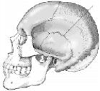
A) coronal.
B) squamosal.
C) sagittal.
D) lambdoidal.
F) B) and C)
Correct Answer

verified
Correct Answer
verified
Multiple Choice
What projection (method) is demonstrated in the image below used to evaluate the cranium?
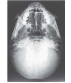
A) PA axial (Caldwell)
B) AP axial (Towne)
C) PA axial (Haas)
D) SMV (Shüller)
F) B) and C)
Correct Answer

verified
Correct Answer
verified
Multiple Choice
Which of the following is true regarding the lateral projection of the skull? 1) The midsagittal plane of the head is parallel to the image receptor. 2) The interpupillary line is perpendicular to the image receptor. 3) The mentomeatal line is parallel with the bottom edge of the image receptor.
A) 1 and 2
B) 1 and 3
C) 2 and 3
D) 1, 2, and 3
F) A) and B)
Correct Answer

verified
Correct Answer
verified
Multiple Choice
The opening into the apex of the orbit for the transmission of the optic nerve and ophthalmic artery is called the:
A) optic canal.
B) foramen.
C) foramina ovale.
D) foramina rotundum.
F) B) and C)
Correct Answer

verified
Correct Answer
verified
Multiple Choice
The suture located between the occipital bone and the parietal bones is the:
A) lambdoidal.
B) squamosal.
C) sagittal.
D) corona.
F) All of the above
Correct Answer

verified
Correct Answer
verified
Multiple Choice
What is the central-ray angulation for the SMV projection?
A) 0 degrees
B) 5 degrees caudad
C) 5 degrees cephalad
D) 5 to 7 degrees cephalad
F) A) and B)
Correct Answer

verified
Correct Answer
verified
Multiple Choice
The axiolateral oblique projection is used to demonstrate the mandible. How is the head positioned to demonstrate the ramus of the mandible?
A) True lateral
B) 30 degrees toward the IR
C) 45 degrees toward the IR
D) 30 to 45 degrees toward the IR
F) C) and D)
Correct Answer

verified
Correct Answer
verified
Multiple Choice
What is the projection (method) demonstrated in the image below used to evaluate the cranium?
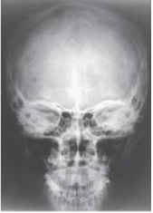
A) Parietoacanthial (Waters)
B) PA axial (Caldwell)
C) AP axial (Towne)
D) PA
F) None of the above
Correct Answer

verified
Correct Answer
verified
Multiple Choice
Which of the following is placed perpendicular to the image receptor for the acanthoparietal projection (reverse Waters method) of the facial bones?
A) Mentomeatal line
B) Infraorbitomeatal line
C) Acanthiomeatal line
D) Glabellomeatal line
F) A) and B)
Correct Answer

verified
Correct Answer
verified
Multiple Choice
What projection and anatomy is demonstrated in the image below?
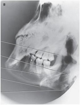
A) AP axial of the TMJs
B) PA axial of the mandibular rami
C) PA of the mandibular body
D) Axiolateral oblique of the mandibular body
F) A) and B)
Correct Answer

verified
Correct Answer
verified
Multiple Choice
The parietoacanthial projection (Waters method) of the sinuses requires the orbitomeatal line to be placed how many degrees from the plane of the image receptor?
A) 20 degrees
B) 27 degrees
C) 30 degrees
D) 37 degrees
F) A) and C)
Correct Answer

verified
Correct Answer
verified
Multiple Choice
What type of joint is the TMJ?
A) Synovial-hinge
B) Synovial-gliding
C) Synovial-ellipsoidal
D) Synovial-hinge and gliding
F) All of the above
Correct Answer

verified
Correct Answer
verified
Multiple Choice
Which two facial bones form the roof of the mouth? 1) Maxillae 2) Vomer 3) Palatine bones
A) 1 and 2
B) 1 and 3
C) 2 and 3
D) 1, 2, and 3
F) B) and C)
Correct Answer

verified
Correct Answer
verified
Multiple Choice
Which line should be placed parallel to the plane of the image receptor for the SMV projection of the cranial base?
A) Acanthiomeatal line
B) Orbitomeatal line
C) Infraorbitomeatal line
D) Mentomeatal line
F) C) and D)
Correct Answer

verified
Correct Answer
verified
Multiple Choice
The midsagittal plane of the head is rotated how many degrees for the axiolateral oblique projection of the TMJ?
A) 0 degrees toward the IR
B) 15 degrees toward the IR
C) 0 degrees away from the IR
D) 15 degrees away from the IR
F) C) and D)
Correct Answer

verified
Correct Answer
verified
Multiple Choice
The part of the sphenoid bone identified in the figure below is the:
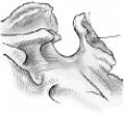
A) clivus.
B) clinoid processes.
C) sella turcica.
D) dorsum sellae.
F) A) and B)
Correct Answer

verified
Correct Answer
verified
Multiple Choice
The posterior half of the base of the skull is formed by which bone?
A) Temporal
B) Sphenoid
C) Occipital
D) Parietal
F) B) and C)
Correct Answer

verified
Correct Answer
verified
Multiple Choice
What structure is labeled as letter B in the image below used to evaluate the cranium?
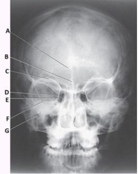
A) Acanthion
B) Crista galli
C) Cribriform plate
D) Petrous ridge
F) A) and D)
Correct Answer

verified
Correct Answer
verified
Showing 21 - 40 of 196
Related Exams